Retinal detachment is an eye condition which occurs when the retina detaches or lifts from the retinal pigment epithelium (RPE). The retina is a thin layer of tissue at the back of the eye, while the retinal pigment epithelium is the layer beneath it that supports its function. When this separation happens, the retina cannot receive the nourishment it needs. As a result, it cannot function properly, leading to changes in vision. If not treated promptly, it can result in permanent loss of vision in the affected eye.
Types of retinal detachment
There are three primary types of retinal detachment.
1. Rhegmatogenous retinal detachment
This is the most common type and it occurs when a tear or hole forms in the retina. Vitreous humour, a gel-like fluid, consequently passes through the tear and collects underneath the retina, causing it to separate from the RPE.
2. Tractional retinal detachment
This occurs when scar tissue on the surface of the retina contracts, pulling away from the RPE. This pulling (traction) parts the retina from its supporting layer. This type of retinal detachment is more common among people with conditions like diabetes.
3. Exudative (serous) retinal detachment
In this type, fluid builds up beneath the retina without a tear. This could be due to inflammation, injury or certain eye conditions.
Causes of retinal detachment
Some of the common causes of retinal detachment include:
Ageing: With age, the gel inside the eye (vitreous) shrinks and can pull on the retina, which is known as Posterior vitreous detachment (PVD). Sometimes this pull creates a tear, which can lead to detachment.
Eye injury: A blow or trauma to the eye can damage the retina directly or cause it to tear.
Myopia (short-sightedness): People with high myopia often have thinner retinas, making their retinas more vulnerable to tearing.
Previous eye surgery: Eye surgeries like cataract removal can slightly increase the risk of retinal tears.
Other eye conditions: Certain conditions, such as diabetic retinopathy and lattice degeneration or inflammation conditions, such as uveitis, that affect the retina, are more likely to cause retinal detachment.
Hereditary factors: A family history of retinal detachment or certain inherited eye conditions can also raise the risk of getting this eye condition.
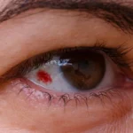
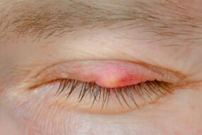
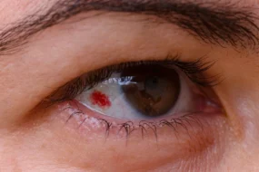
Symptoms of retinal detachment
Common signs and symptoms include:
• Photopsia (flashes of light which appear as streaks of light or flickers)
• A sudden increase in floaters (small dark dots, threads or cobweb-like shapes)
• Blurry or distorted vision
• A shadow, curtain or dark veil effect across your vision field
If you notice any of these retinal detachment symptoms, seek medical attention immediately. Early treatment is essential to help prevent permanent vision loss.
Diagnosis of retinal detachment
Retinal detachment is diagnosed through a thorough eye examination by an optometrist or an ophthalmologist. They will dilate your pupils with eye drops so the back of your eye can be examined clearly using an ophthalmoscope. They will then use a slit‑lamp microscope with a bright light to examine your eye while you look in various directions, checking for any tears, holes or areas of separation.
If the retina cannot be seen clearly, for example, due to bleeding inside the eye, an ultrasound scan may be used to assess the internal structure.
Typically, both eyes will be examined, even if symptoms are only present in one. You may also be asked to attend a follow-up appointment to ensure no delayed retinal tears develop.
Treatment for retinal detachment
Treatment for a detached retina depends on the stage and severity of the condition. In most cases, surgery is required to reattach the retina and restore its position at the back of the eye.
There are three main surgical approaches:
Vitrectomy: This procedure involves removing the gel (vitreous) from inside the eye. Any retinal tears present are repaired and a gas bubble or silicone oil is placed inside the eye to hold the retina in its correct position while it heals.
Scleral buckle surgery: A soft silicone band is placed around the sclera (the white part of the eye), gently supporting the eye wall and helping the retina settle back into place. The band is usually left in the eye permanently and is not visible.
Pneumatic retinopexy: A small gas bubble is injected into the eye to press the retina back into position. The retinal tear is then sealed to allow healing.
The most suitable treatment option depends on the type and extent of the detachment. Your ophthalmologist will recommend the most suitable procedure based on your condition.
Frequently Asked Questions
What are the benefits of retinal detachment surgery?
The primary benefit is to repair the detached retina and prevent the condition from worsening. Prompt treatment can improve chances of preserving vision.
What complications can occur after retinal detachment surgery?
Complications are quite uncommon, but as with any surgery , they can occur. Possible complications of retinal detachment surgery include cataract formation, inflammation, double vision, allergy, changes in eye pressure and eye infections. Sometimes additional surgery may be necessary.
Can you wear contact lenses after retinal detachment surgery?
Yes, you can wear contact lenses again once the eye has healed and your ophthalmologist confirms it is safe to wear them. Recovery time varies depending on the procedure.
Disclaimer: The advice in this article is for informational purposes only and does not replace medical care or an in-person check-up. Please check with an eye care professional before purchasing any products or remedies. For information on our article review process, please refer to our Editorial Policy.

Tina Patel is a qualified Contact Lens Optician at Feel Good Contacts with over 25 years of experience in the optical industry. She is a member of ABDO and registered with the GOC. Tina holds a BSc. (Hons) in Optical Management from Anglia Ruskin University and qualified as a Dispensing Optician in 2002. In 2007, she further specialised by completing advanced training in contact lenses, gaining her qualification as a Contact Lens Optician. She now shares her clinical expertise through teaching at City, University of London, guiding future opticians.